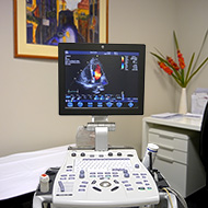
Echocardiography
Echocardiography (also called Echo-Doppler or Cardiac Ultrasound) is a diagnostic test that uses high frequency sound waves (ultrasound) to provide an image of the heart. Valuable information about the function of the pumping chambers (ventricles), the heart valves, and blood flow through the heart, is obtained by placing an echo probe onto the chest wall.
Echocardiography may be requested to investigate problems that your doctor thinks could be due to a cardiac condition. Patients with a variety of symptoms might benefit with Echocardiography including: shortness of breath, dizziness and blackouts, chest pain, palpitations, loss of energy. Sometimes, echocardiography is performed to assess a heart murmur or to provide a baseline prior to treatments that might affect the heart (e.g. chemotherapy).
Understanding the testing procedure
The test is performed within our Consulting Rooms and does not require any special preparation. The test takes 30-40 minutes to complete but, sometimes, a longer time might be required. One of our expert Echo Technologists will perform the test and will spend as much time as necessary to acquire the best possible images.
Echocardiography is safe and painless; the ultrasound used during this test has never been shown to have any harmful effects and is perfectly safe for expectant mothers. However, you might experience a small amount of discomfort due to pressing the probe onto your chest. The test is performed with you lying on your left side, on a couch specially designed for this purpose.
Once the images have been acquired, a cardiologist views them and the findings formally reported to your doctor.
